MRI Scan for Mesothelioma: Imaging & Diagnosis Guide
Magnetic resonance imaging (MRI) plays a crucial role in mesothelioma diagnosis and treatment planning. Unlike CT scans that use radiation, MRI creates detailed images of soft tissues using magnetic fields. This guide explains how MRI is used to detect mesothelioma, assess disease extent, and help oncologists plan personalized treatment strategies.
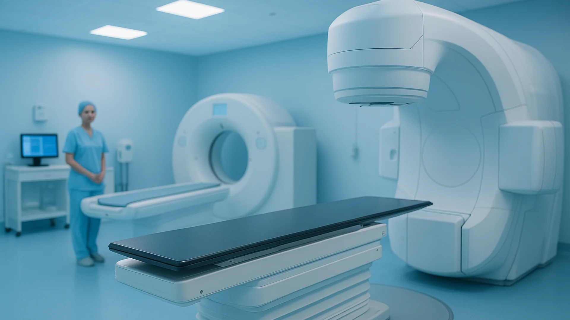
Table of Contents
What is an MRI Scan?
Magnetic resonance imaging (MRI) is a non-invasive diagnostic imaging technique that uses a powerful magnetic field and radio waves to create detailed images of the body's internal structures. Unlike X-rays and CT scans, MRI does not use ionizing radiation, making it a safer option for patients requiring multiple scans or those concerned about radiation exposure.
The MRI machine produces a strong magnetic field that aligns hydrogen atoms in your body. Radio waves are then applied, causing these atoms to emit signals that are detected and processed by the scanner to create cross-sectional images. These images can be viewed in multiple planes (axial, sagittal, and coronal) to provide a comprehensive view of the area being examined.
MRI is particularly valuable for imaging soft tissues, including organs, muscles, fat, and blood vessels. The machine is large and cylindrical, and the scanning process is completely painless, though it produces loud knocking sounds during operation. MRI scans for mesothelioma typically focus on the chest (for pleural mesothelioma) or abdomen (for peritoneal mesothelioma).
MRI in Mesothelioma Diagnosis
MRI plays an important supporting role in mesothelioma diagnosis, though it is typically not the first imaging test ordered. While CT scans are often used initially to detect suspicious pleural thickening or masses, MRI provides superior soft tissue visualization and is particularly useful for:
- Assessing pleural thickening: MRI can distinguish between benign pleural thickening and malignant tumor involvement
- Detecting diaphragmatic invasion: MRI is excellent for visualizing whether the tumor has invaded the diaphragm, a critical factor for surgical planning
- Evaluating chest wall involvement: Detailed images help determine if the cancer has spread to the chest wall, ribs, or underlying muscles
- Assessing pericardial involvement: For pericardial mesothelioma, MRI can detect involvement of the heart sac
- Determining surgical resectability: High-quality MRI images help surgeons plan operations and determine if the tumor can be completely removed
MRI is also valuable for monitoring disease progression during treatment. Comparing sequential MRI scans allows your oncology team to assess how well treatment is working and detect any changes in tumor size or location.
Detection and Staging
In pleural mesothelioma, MRI can detect abnormal pleural thickening that may indicate cancer. The imaging technique excels at showing the relationship between the tumor and surrounding anatomical structures. This spatial relationship is critical for determining disease stage and treatment feasibility.
For peritoneal mesothelioma, MRI can identify tumor nodules in the peritoneum and assess their distribution throughout the abdomen. It can also detect ascites (fluid accumulation) and involvement of abdominal organs. This information is essential for determining candidacy for cytoreductive surgery with heated intraperitoneal chemotherapy (HIPEC).
MRI is particularly valuable for detecting early-stage disease and identifying subtle metastases that might be missed on CT. The superior soft tissue contrast of MRI makes it ideal for precise staging, which directly impacts treatment recommendations and prognosis.
Advanced MRI techniques such as diffusion-weighted imaging (DWI) and dynamic contrast-enhanced imaging can provide additional information about tumor characteristics and blood flow patterns, helping differentiate between benign and malignant lesions.
MRI vs. Other Imaging Tests
Mesothelioma diagnosis typically involves multiple imaging modalities, each with distinct advantages. Understanding how MRI compares to other imaging tests can help you better understand your diagnostic workup.
CT Scan vs. MRI: CT scans are usually the first imaging test for mesothelioma because they are faster, widely available, and excellent for detecting pleural thickening and masses. However, MRI provides superior soft tissue contrast and does not expose patients to radiation. CT is better for detecting lung nodules, while MRI is superior for assessing invasion into surrounding structures.
Chest X-ray vs. MRI: Chest X-rays are often the initial screening tool and are inexpensive and quick, but they have limited detail for soft tissue visualization. MRI provides much more detailed anatomical information but takes longer and is more costly.
PET Scan vs. MRI: PET scans use radioactive tracers to identify areas of high metabolic activity, making them useful for detecting metastatic disease. MRI is better for precise anatomical staging and local tumor assessment. These tests are often used together for comprehensive staging.
Most mesothelioma patients undergo a combination of imaging tests—typically CT for initial detection, MRI for detailed staging and surgical planning, and PET for metastatic evaluation. Your doctor will determine which imaging tests are appropriate for your specific situation.
The MRI Procedure
Understanding what to expect during an MRI scan can help reduce anxiety and ensure you're well-prepared. A typical mesothelioma MRI procedure takes 30 to 45 minutes from start to finish, though the actual scanning may take 20 to 30 minutes.
Before the scan: You'll be asked to change into a hospital gown and remove all metal objects, including jewelry, glasses, watches, and hearing aids. The MRI technologist will review your medical history and ask about any metal implants, including pacemakers, surgical clips, or metallic foreign bodies.
During the scan: You'll be positioned on a moving table that slides into the cylindrical MRI machine. The machine produces a strong magnetic field and loud banging or knocking sounds. Some facilities provide earplugs or headphones. You'll need to remain still during scanning to ensure clear images. The technologist can see and hear you throughout the procedure.
Contrast injection: For mesothelioma imaging, gadolinium-based contrast is usually injected intravenously to enhance visualization of tumors and vascular structures. This happens midway through the scan and helps differentiate between tumor and non-tumor tissue.
Breathing instructions: Your technologist may ask you to hold your breath for brief periods during scanning to minimize motion artifacts. Following these instructions carefully is important for obtaining high-quality images.
Safety Considerations
MRI is generally a very safe imaging technique with minimal health risks. Unlike CT and X-ray imaging, MRI uses no ionizing radiation, eliminating radiation exposure concerns. However, there are important safety considerations to discuss with your healthcare team.
Metal implants and devices: The strong magnetic field can affect or damage certain metallic implants. Patients with pacemakers, implantable defibrillators, or certain other medical devices may not be able to undergo MRI. Non-metallic alternatives for these devices are increasingly available, so inform your doctor about any implants before scheduling an MRI.
Metallic foreign bodies: Small metal fragments in the eye or embedded in tissue can be dangerous in an MRI machine. Always disclose any history of metal work or eye injury.
Claustrophobia: Some patients experience anxiety or claustrophobia in the confined MRI machine. Inform your doctor if you have significant anxiety; open-sided MRI machines are available at some facilities.
Gadolinium contrast: While gadolinium is generally safe, patients with severe kidney disease (GFR less than 30) have a slightly increased risk of nephrogenic systemic fibrosis. Your kidney function will be assessed before contrast administration.
Preparing for Your MRI
Proper preparation ensures your MRI scan goes smoothly and produces high-quality diagnostic images. Follow these preparation guidelines:
- Arrive 15 minutes early to complete check-in and paperwork
- Remove all metal objects: jewelry, watches, glasses, hearing aids, and hairpins
- Wear comfortable, metal-free clothing or change into a hospital gown
- Inform the technologist about all metal implants and medical devices
- If you have tattoos, mention them to your technologist (most are safe, but some older tattoos contain metal)
- Eat and drink normally unless instructed otherwise
- Take medications as directed, unless your doctor instructs otherwise
- If you're receiving contrast, ensure your kidneys are functioning well (labs may be needed)
- Use the restroom before the scan
- Plan for someone to drive you home if you received contrast agent
After Your MRI Scan
After your MRI is complete, there are no restrictions on activity. You can drive yourself home, return to work, and resume all normal activities immediately. If gadolinium contrast was used, drink plenty of water to help flush the contrast from your body.
Your MRI images will be reviewed by a radiologist, who will prepare a detailed report. The report will be sent to your oncologist or referring physician, typically within 24 to 48 hours. Your doctor will discuss the results with you and explain what the findings mean for your diagnosis, staging, and treatment plan.
If you have any questions about your MRI results or need clarification on what they mean, don't hesitate to ask your medical team. Understanding your imaging results is an important part of being an informed patient in your cancer care.
Medically Reviewed
This article was reviewed by Dr. James Mitchell, MD, a board-certified radiologist specializing in oncologic imaging. Last reviewed: March 22, 2026.
Know Your Legal Rights
If you've been diagnosed with mesothelioma, you may be entitled to compensation from negligent employers or asbestos manufacturers. A mesothelioma attorney can help you understand your options.
Get Legal InformationFrequently Asked Questions
- How is an MRI scan different from a CT scan?
- MRI uses magnetic fields instead of radiation. CT scans are faster and better for lung imaging, while MRI provides superior soft tissue visualization without radiation exposure.
- Can I eat before an MRI?
- Yes, you can eat and drink normally before an MRI unless your doctor instructs otherwise. If contrast is planned, eat a light meal beforehand.
- What if I'm claustrophobic?
- Inform your doctor and MRI facility about claustrophobia. Open-sided MRI machines are available at some centers, or sedation may be an option.
- How long before I get my results?
- A radiologist will review your MRI within 24 to 48 hours. Your doctor will discuss the results with you and explain their significance for your treatment plan.
- Will the MRI show all the information needed for treatment planning?
- MRI provides valuable information about tumor extent and involvement of surrounding structures. However, your oncologist may recommend additional tests such as CT or PET to ensure comprehensive staging.